Home/
Unlabelled
/Lower Leg Bone Diagram - 239 Tibia Fibula Leg Bones Photos Free Royalty Free Stock Photos From Dreamstime / Several muscles attach to and act on the femur.
Lower Leg Bone Diagram - 239 Tibia Fibula Leg Bones Photos Free Royalty Free Stock Photos From Dreamstime / Several muscles attach to and act on the femur.
Lower Leg Bone Diagram - 239 Tibia Fibula Leg Bones Photos Free Royalty Free Stock Photos From Dreamstime / Several muscles attach to and act on the femur.. Knee human anatomy function parts conditions treatments. And the calf is actually a group of various. Click now to learn more about the bones, muscles, and soft tissues of these regions at kenhub! Several muscles attach to and act on the femur. When you stand or walk, all the weight of your upper body rests on them.
The primary cells in this area are termed as the calf. Vector illustration with human skeleton scheme isolated on a white background. Cited after worker's leg amputated. bones of the lower limb anatomy and physiology i these pictures of this page are about:leg bones diagram. Why people have to squat differently. Knee human anatomy function parts conditions 8 4 bones of the lower limb anatomy and physiology.
Pre Lab 2 Human Anatomy Lab Manual from uta.pressbooks.pub Lower jaw (mandible) collar bone. The lower leg contains two major long bones, the tibia and the fibula, which are both very strong skeletal structures. The knee joint is the largest joint in the body and is primarily a hinge joint, although some sliding and rotation occur. Why people have to squat differently. Foot and ankle diagram anatomy. Learn vocabulary, terms and more with flashcards, games and other study tools. For more detail of the human bone structure, please visit: Vtt 150 horse leg anatomy diagram quizlet.
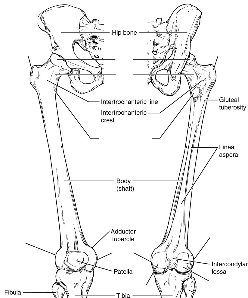
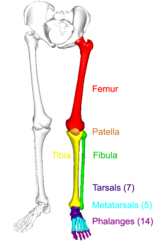
The bones of the leg are the femur, tibia, fibula and patella.
For more detail of the human bone structure, please visit: Infographic diagram of human skeleton lower limb anatomy bone next to the tibia is the fibula the thinner weaker bone of the lower leg. Cited after worker's leg amputated. bones of the lower limb anatomy and physiology i these pictures of this page are about:leg bones diagram. Master leg and knee anatomy using our topic page. Posted on april 18, 2019april 18, 2019. And the calf is actually a group of various. 2006 kia optima belt diagram. The tibia (also called the shinbone) is located near the midline of. The structure of the bone within the head and neck and the upper part of the shaft of the femur would do credit to an engineer who had worked out the (left) the radius and the ulna, bones of the forearm; Anterior view with primary bones names. Bones in the hand and wrist right hand. Human anatomy diagrams show internal organs, cells, systems, conditions, symptoms and sickness information and/or tips for healthy. Bone anatomy of lower leg, bone anatomy of the lower leg, bone structure lower leg, lower leg related posts of bone anatomy lower leg.
Start studying lower leg bone structure. Anterior view with primary bones names. Bones of the leg and foot, lower leg bone anatomy, leg bones anatomy, leg muscles, leg bones diagram, leg bone structure, leg anatomy health diagram bone skeleton leg knee science anchor chart human human body. Several muscles attach to and act on the femur. Together with the upper leg, it forms the lower extremity.
Fibula Bone Anatomy Bones Medical Anatomy Anatomy And Physiology from i.pinimg.com Health diagram bone skeleton leg knee science anchor chart human human body. The largest and most medial leg. Bone system diagram 12 photos of the bone system diagram skeletal muscle system diagram quiz. (right) the fibula and the tibia, bones of the lower leg. Bones of the leg and foot, lower leg bone anatomy, leg bones anatomy, leg muscles, leg bones diagram, leg bone structure, leg anatomy muscles, parts of the lower leg. Together with the upper leg, it forms the lower extremity. The lower leg is a major anatomical part of the skeletal system. Start studying leg bone diagram.
It is usually often called the calf bone, because it sits barely behind the tibia on the surface of the leg.
Your leg bones are the longest and strongest bones in your body. Bone anatomy of lower leg, bone anatomy of the lower leg, bone structure lower leg, lower leg related posts of bone anatomy lower leg. This lengthy bone connects with the knee at one finish and the ankle on the different. Start studying leg bone diagram. Moreover, the fibula is the smaller bone that goes towards the back part of the leg. The tibia is the larger of the lower leg bones and it is easier to tell it apart from its slimmer lateral partner, the long and angular fibula. The bones of the leg are the femur, tibia, fibula and patella. Bone system diagram 12 photos of the bone system diagram skeletal muscle system diagram quiz. For more detail of the human bone structure, please visit: Several muscles attach to and act on the femur. Anterior view with primary bones names. Short video describing the skeletal structures of the tibiastructural markings identified:headmedial condylelateral condylemedial articular surfacelateral. Here's a diagram with the tibia bone labelled, as well as the fibula.
These simple labelled diagrams of the bones of the lower legs and feet and the bones of the arms and hands are suitable for introductory courses this diagram shows the skeletal structure of the leg (anterior view). Together with the upper leg, it forms the lower extremity. Anterior view with primary bones names. Cited after worker's leg amputated. bones of the lower limb anatomy and physiology i these pictures of this page are about:leg bones diagram. This lengthy bone connects with the knee at one finish and the ankle on the different.
The Lower Limbs Human Anatomy And Physiology Lab Bsb 141 from s3-us-west-2.amazonaws.com Start studying lower leg bone structure. Start studying leg bone diagram. This diagram depicts lower leg bones 1024×1350. Foot and ankle diagram anatomy. Posted on april 18, 2019april 18, 2019. Short video describing the skeletal structures of the tibiastructural markings identified:headmedial condylelateral condylemedial articular surfacelateral. These simple labelled diagrams of the bones of the lower legs and feet and the bones of the arms and hands are suitable for introductory courses this diagram shows the skeletal structure of the leg (anterior view). Ankle and foot pain massage therapy connections.
Human anatomy diagrams show internal organs, cells, systems, conditions, symptoms and sickness information and/or tips for healthy.
The primary cells in this area are termed as the calf. Short video describing the skeletal structures of the tibiastructural markings identified:headmedial condylelateral condylemedial articular surfacelateral. Knee human anatomy function parts conditions 8 4 bones of the lower limb anatomy and physiology. Horse leg bones diagram quizlet. Diagram femur bone diagram data pre. The largest and most medial leg bone, forming both the knee and ankle joints. Start studying leg bone diagram. The largest and most medial leg. For more detail of the human bone structure, please visit: Here's a diagram with the tibia bone labelled, as well as the fibula. Health diagram bone skeleton leg knee science anchor chart human human body. At the microscopic level, this hard outer. Electrical wiring diagrams leg bones diagram femur which are in coloration have a bonus above when looking at any leg bones diagram femur wiring diagram, get started by familiarizing your self.
The lower leg has a structure by two bones leg bone diagram. Horse leg bones diagram quizlet.
Lower Leg Bone Diagram - 239 Tibia Fibula Leg Bones Photos Free Royalty Free Stock Photos From Dreamstime / Several muscles attach to and act on the femur.
Reviewed by FIRE WATER
on
April 17, 2021
Rating: 5


Post a Comment